Basis of CT Imaging
Learning Objectives
- Understand the basic principles of CT scanning
- Understand the basic principles of CT image formation
- Understand the source of typical artefacts in CT imaging
What is Computed Tomography?
Computed Tomography (CT) is a radiography technique in which a three-dimensional image of a body structure is constructed by computer from a series of plane cross-sectional images made along an axis.
The word tomography is derived from Ancient Greek τόμος tomos, “slice, section” and γράφω graphō, “to represent, study”.
CT is a very widely used general diagnostic radiographic technique. However compared to a planar x-ray instead of just taking one image it takes as series of projections around an axis forming many different slices. More clinically useful information can be received with a CT scan but at the cost of giving the patient a higher dose.
Planar X-Rays vs CT
Planar radiography renders a 3D volume onto a 2D image. Conventional planar skull X-rays (SXR) were traditionally poor for head images due to lots of overlapping structures and skull dominating the projection making it hard to see any other information in the head. However with a sliced CT image one can clearly see the the ventricles in the brain. There are more problems with planar x-rays as well:
- Contrast in planar x-rays is very good for bone due to the differences in attenuation but is very hard to differentiate between softer tissues.
- Spatial relation as planar x-rays form a 2D image it is often hard to see where objects are spaced and where they lie in relation to each other. There is a loss of depth. Whereas in CT you can see small differences a lot clearer.
Tomographic Reconstruction
Is where different attenuation (or CT number) values are mapped out in order to form an image. It was developed by Johann Radon in 1917 and is related to Fourier Transforms.
Radon’s Theorem
Given an infinite number of one-dimensional projections taken at an infinite number of angles, you can reconstruct the original object perfectly
Basic Principles of CT Formation
CT Scanner design
The basic components of CT Scanner are:
- X-ray Tube: the source, which projects a fan beam
- X-ray detector: traditionally film, then Computed Radiogrpahy (CR), now Direct Radiography (DR).
- X-ray attenuator: the patient or thing to be x-rayed Motion: means to gather projection data from various angles, usually the x-ray tube and detector are connected together on slip rings.
First Generation CT Scanner
The first generation of CT Scanner had the following characteristics:
- Narrow pencil beam
- Single detector
- Translational and rotational movement around the patient
- Very Slow it took minutes per slice
Third Generation CT Scanner
The third (current) generation of CT scanner vastly improved upon the previous generations and had the following characteristics:
- Fan beam
- Multiple detectors
- Rotation only and no translation is required
- Much faster and is a fast as 0.5s per rotation
X-ray tube advances
CT is very demanding of X-ray tubes and generators as scans are required instantaneously (up to 700 mA) and sequences can take over 30 seconds. This requires a large heat capacity and fast cooling rates. Mechanical stresses due to tube rotation are very large as well and are over 15G for 0.5s rotation.
Flat Filtration
Filtration in the x-ray tube and housing absorbs low energy X-rays, which contribute to the patient dose but not to the image quality. Equivalent to as much as 13mm aluminium.
Beam shaping filter Bow-Tie
After normal filtration, another filter is present in a CT scanner and known as a bow-tie filter. The edges of the patient to be x-rayed are thinner and therefore have less attenuation. This bow-tie filter lessens the intensity of the x-ray beam at the edges (more intense beam at the centre) so the beam incident at the detectors is more constant and it also removes soft X-rays.
Detectors
These are a critical component as they record the intensity of the incident X-rays sending out a signal. There are many different types including:
- Xenon: Older variant uses pressurised Xenon gas, ionisation.
- Solid Sate: More common for CT imaging. In uses scintillation, photon capture, light-photo-diode-signal.
These detectors have to perform well and be efficient under lots of physical stress
Detector Arrangement
Detectors in 3rd generation scanners are arranged in an arc around the patient. There are approximately 600 to 900 elements in a detector bank which allows for good spatial information. Both the tube and the detectors rotate around the patient.
Helical CT Scanning
Helical Acquisition
To try and reduce dose and make scanning even faster, the table also moves while X-Ray tube and detector rotate during acquisition. A problem with this is that some gaps can form in the images of the patient, however these can be filled in with interpolation.
Helical Pitch
Is the speed of the table movement through the gantry defines the spacing of the helices.
EQUATION
$ Helical \ Pitch = \frac{Table \ Travel \ per \ Rotation}{X-ray \ beam \ Width} $
For Example: An increase in pitch increases the movement of the table, decreases the amount of time required however leads to more gaps between the images.
Helical Image Reconstruction
To reconstruct the data as normal, the CT scanner uses a combination of data 180° each side of a recon (reconstruction) position and interpolation. Interpolation averages data on either side of the reconstruction position to estimate the projection data at that point.
As a result, a interpolated helical scan is able to reduce artefacts due to changing structure in z-axis when moving the table. For any set reconstruction position, only one scan projection will be available at that point.
Note: Data at 180° at either side of the recon position is more commonly used than 360° on one side as z-axis interpolation distances are shorter. Also 180° interpolator makes use of the opposite (Anterior-Posterior & Positive-Anterior) views producing a second complementary spiral for interpolation
Advantages
A few advantages of helical scanning are:
- Speed: as there is no need to pause between scans for table movement, pitches greater than 1 are possible, pitches less than 1 are possible, reduced patient movement.
- More information but not exact!, arbitrary image position, can reconstruct overlapping images.
Disadvantages
A disadvantage is the broadening of the slice profile, however this can be overcome using a 180° interpolator at the expense of image noise.
Image reconstruction
The objective of CT image reconstruction is to determine how much attenuation of the narrow x-ray beam occurs in each voxel of the reconstruction matrix. These calculated attenuation values are then represented as grey levels in the 2D image of the slice.
Linear Attenuation Coefficient µ
All tissues or material has a linear attenuation coefficient which is summed along the path between the tube and the detectors, as well as varying with energy.
- High µ is for dense, high atomic number (z) materials. As the attenuation of X-rays is high, this gives a low signal to detectors
- Low µ is for low density, low atomic number (z) materials. Lower attenuation, higher signal to detectors
Note: µ relates to z (atomic number), ρ (physical density) and E (X-ray energy)
What is a CT image?
A map or array of picture elements (pixels) presenting a grey scale (pixel value) relating to the stored value which is the calculated result of the tomographic reconstruction of projection data relating to the attenuation in a volume cell (Voxel) in a patient. Each 2D pixel in a CT image represents average attenuation within a 3D voxel.
Data Acquisition
Many attenuation measurements are taken, with a sample at each detector position generating a profile. This gives view of a patient at one orientation; a projection. A number of projections are collected from views all around the patient. Different manufacturers have different combinations of detectors.
Filtered Back projection FBP
Back projection is a mathematical function that is applied to the attenuation data that you find, which reverses the process of measurement of projection data to reconstruct an image. Each projection is ‘smeared back’ across the reconstructed image. Consider each projection as an intensity map, where white is high attenuation (something ‘hard’) and dark is low attenuation (nothing there).
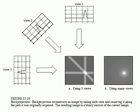
However the output back projection trans-axial image is blurry. The projection data needs to be processed before reconstruction. Kernels (mathematical filters) can be applied for different diagnostic purposes. Smoothing for viewing soft tissue and sharpening for high resolution imaging. This post-processing in combination with back-projection is known as filtered back projection.
CT Number HU
A normalised attenuation number using fixed reference points of water & air.
$ CT = \frac{\mu_{tissue} - \mu_{water}}{\mu_{water}} \times 1000 $
Hounsfield units are the standard units for CT number in medical imaging with water at 1000 HU and air at -1000 HU.
We can change the appearance of the image by varying the Window Level (WL) and Window Width (WW). This spreads a small range of CT numbers over a large range of grayscale values, making it easy to detect very small changes in CT number.
- Window Level (WL) is the CT number of mid-grey.
- Window Width (WW) is the number of HU from black to white.
CT imaging artefacts
Ring artefacts
This occurs in 3rd generation CT scanners. If one detector is out of calibration with the other detectors, this consistently gives erroneous readings at each projection. A circular artefact is presented.

Partial Volume effects
If an object is continuous is the z-axis the CT number is not affected by the z-sensitivity. If an object varies in z-axis (especially using helical scanning), the ‘partial volume effect’ will alter CT number. To solve this problem the pitch can be reduced.
Beam Hardening artefacts
As an x-ray beam passes through a material is becomes more attenuated and becomes ‘harder’ the further it travels. The peak energy of the x-ray beam starts moving higher up the spectrum, so becomes more penetrating and more intense at the detectors. This various artefacts to appear:
- Cupping: the central x-rays are hardened due to a decrease in attenuation rate compared to the edges. The beam is therefore more intense at the detectors.
- Streaks and dark bands: appear between dense objects. As the beam goes through both objects at some projections and one object for others. Beam passing through only one object is hardened less than those passing through both objects.
Metal artefacts
These are caused when density of material is beyond the normal range of a scanner computer (incomplete attenuation profiles). It is compounded by beam hardening, partial volume and aliasing. Filters can be applied to reduce the metal artefacts, but ideally metal objects are removed however this is not possible with implants!
Motion artefacts
If the object moves while the scanning takes place, misregistration artefacts appear as shading or streaking. To prevent this, the CT operator will tell a patient to hold their breath will scanning, to minimise any movement due to breathing.
Written by Tobias Whetton